The imaging techniques , in suitable parameters , provide the connection between the cell therapies and the clinical procedure . The longitudinal monitoring provides information about the functioning mechanism of the reprogrammed cells in the in vivo environment . The review presents the imaging techniques that are able to explain the cause for the limited restoration process of the cell therapies in the cardiac tissue .
The human body contains approximately 3 , 7 × 10¹³ cells . The heart contains 6 × 10⁹ cells , in 300 grams ( figure 1 ) . The damage of the heart does not lead automatically to the regeneration of the full functional capacity . The scar formation and the inflammatory signals may not be enough to have a full regeneration of the cardiac tissue . The regenerative medicine enhances the healing mechanisms of the body with the cell therapy .
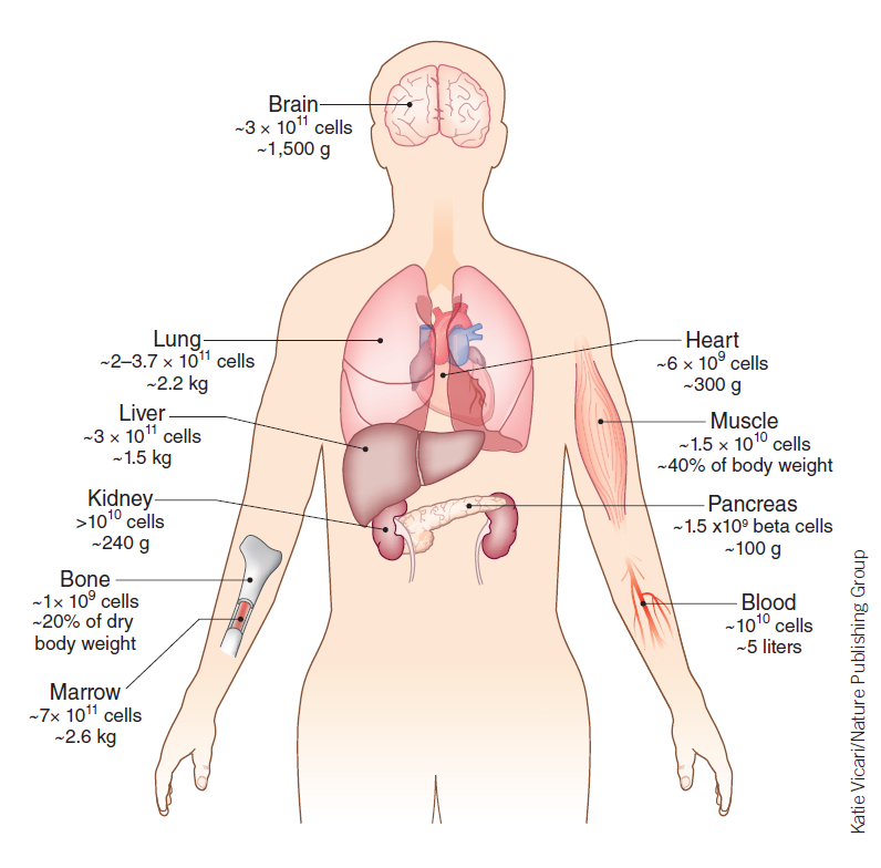
Figure 1 . The number of cells in the parts of the human body .
Copyright ( 2014 ) Katie Vicari / Nature Publishing Group
The minimum number of injected cells that is detectable by the imaging techniques is in the range of 104 - 106 cells . The heart contains 2 × 10⁷ cells in one gram of tissue , in the left ventricle . The fused positron emission tomography ( PET ) - computed tomography ( CT ) image of the porcine heart is an example for the technique ( figure 2 ) . The long arrows point to the trajectory of the thoracotomy . T is the inserted tube . The short arrows point to the 108 human mesenchymal stem cells , injected in the left ventricle ( LV ) . The %ID / g is the percentage of the uptake by the injected human cells in one gram of porcine cardiac tissue .

Figure 2 . The PET - CT imaging of the expression for the reporter gene in the porcine myocardium .
Copyright ( 2009 ) RSNA
The results of a porcine model for the myocardial infarction identifies as the time for the maximum proliferation at an interval of 33 - 35 days after the injection of the mesenchymal stem cells in the myocardium .
The injected cells are not tracked in vivo if they are not labelled in vitro . The nanoparticles , as the exogeneous label , provide a strong contrast in a short acquisition time , but it is unreliable in the long - term monitoring . The incorporation process of reporter genes labels indirectly the target cell . The reporter gene in the nucleus creates reporter proteins secreted in the cytoplasm . The imaging technique detects the reporter proteins . The ideal cell label stays inside the target cells for a long time , is non - toxic , is in a concentration that is stoichiometric related to the number of target cells , and clears the system rapidly after the apoptosis of the target cells .
The challenges of the imaging techniques are the label dilution , and the cell tracking . The label dilution should remain inside the tracked cells until the apoptosis , but sometimes the dilution transfers to the host cells . The long - term tracking of the implanted cells through the imaging techniques has the challenge to quantify the number of cells from the initial implantation procedure that are alive , and the number of cells that proliferate , in the spatial and the temporal dimension .
The infarcted myocardium is trully regenerated with the aid from the cell therapies when the viable cardiac tissue in the infarct area has the volume increased , the fiber architecture is integer in structure , and the regenerated myocardium has synchronous contraction with the host .
References :
Naumova A . V . , Modo M . , Moore A . , Murry C . E . , Frank J . A . ( 2014 ) “ Clinical imaging in regenerative medicine ” , Nature Biotechnology , 32 ( 8 ) , 804 - 818 .
Willmann J . K . , Paulmurugan R . , Rodriguez - Porcel M . , Stein W . , Brinton T . J . , Connolly A . J . , Nielsen C . H . , Lutz A . M . , Lyons J . , Ikeno F . , Suzuki Y . , Rosenberg J . , Chen I . Y . , Wu J . C . , Yeung A . C . , Yock P . , Robbins R . C . , Gambhir S . S . ( 2009 ) “ Imaging gene expression in human mesenchymal stem cells : from small to large animals ” , Radiology - Radiological Society of North America , 252 ( 1 ) , 117 - 127 .